

Corneal Diseases
The eye's clear front window
At Sutherland Eye Surgeons, we provide specialist treatment of diseases that affect the front of your eye such as pterygia, kerataconus and corneal ulcers.
What are corneal diseases?
The cornea is composed of five distinct layers and serves as the eye's primary refractive surface. When disease affects the cornea, it can cause pain, light sensitivity, and progressive vision loss that cannot be corrected with glasses alone. Common corneal conditions include:
Pterygium is a raised, fleshy, wedge-shaped growth of tissue that starts on the white of the eye and can slowly grow onto the cornea. Often associated with chronic ultraviolet (UV) exposure and dry, dusty environments, it can cause irritation, redness, astigmatism, and blurred vision if it encroaches upon the visual axis.
Keratoconus is a progressive condition in which the cornea thins and bulges into a cone shape, causing irregular astigmatism and significant visual distortion. It typically presents in adolescence or early adulthood and may be associated with allergic eye disease and vigorous eye rubbing.
Corneal ulcers are open sores on the cornea, usually resulting from infection (bacterial, viral, fungal, or parasitic) or severe dry eye. They are often associated with contact lens wear, trauma, or pre existing corneal disease. Corneal ulcers require urgent evaluation, as they can lead to scarring, thinning, and permanent vision loss if not promptly treated.
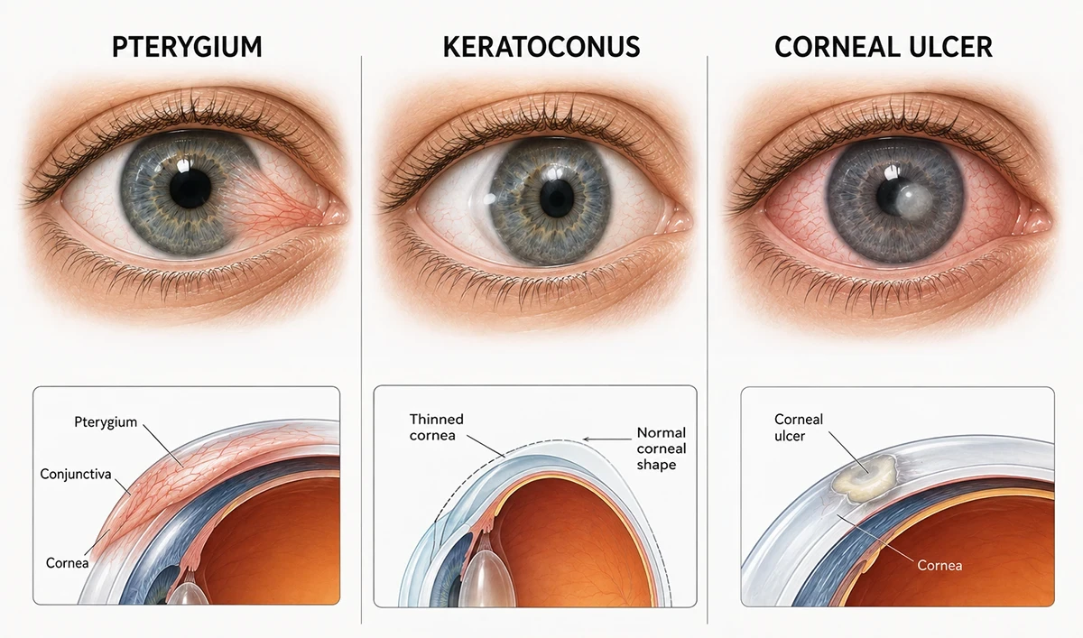
Symptoms of corneal diseases
Pterygium – Redness, irritation, a visible growth on the white of the eye, foreign body sensation, and blurred vision if the growth extends over the pupil or induces astigmatism
Keratoconus – Progressive blurring and distortion of vision, increased sensitivity to light, frequent changes in spectacle prescription, and difficulty with night driving due to glare
Corneal ulcers – Severe eye pain, redness, excessive tearing, light sensitivity, blurred vision, and a visible white or grey spot on the cornea
Assessing corneal diseases
When you visit us at Sutherland Eye Surgeons, some of the tests that we perform may include:
Visual acuity and refraction
Assessment of your best corrected vision and any refractive errorSlit lamp examination
Microscopic examination of the cornea to assess the type, location, and extent of pathology, including the presence of a pterygium, corneal thinning, scarring, or ulcerationCorneal fluorescein staining
The application of a yellow dye that highlights epithelial defects, such as corneal ulcers, under a blue lightCorneal topography
A non invasive imaging technique that maps the corneal surface, essential for diagnosing and monitoring keratoconus, and for guiding the fitting of specialty contact lensesCorneal pachymetry
Measurement of corneal thickness, which is important in assessing the severity of keratoconus and for planning cross linking or transplantationMicrobiological testing
For corneal ulcers, corneal scrapings may be taken for culture and sensitivity to identify the causative organism and guide antibiotic or antifungal therapy
Treatment of corneal diseases
- Pterygium
Mild cases are managed with preservative free lubricating eye drops to reduce irritation, along with UV protection using sunglasses and hats. For larger pterygia threatening vision, causing persistent discomfort, or producing significant astigmatism, surgical excision is indicated. This involves removing the pterygium tissue and performing a conjunctival autograft to cover the bare sclera, significantly reducing the risk of recurrence.
- Keratoconus
Rigid gas permeable (RGP) contact lenses create a smooth optical surface over the irregular cornea, while scleral lenses vault over the entire cornea to provide superior comfort and vision for advanced cases. More severe cases may necessitate referral to a corneal subspecialist for consideration of surgical procedures such as collagen cross linking or corneal transplant surgery.
- Corneal Ulcers
Corneal ulcers require urgent treatment to prevent scarring, perforation, and vision loss. Management typically involves frequent, intensive topical antibiotic or antifungal eye drops based on culture results, along with cycloplegic drops to reduce pain and prevent complications. For deep ulcers or small perforations, tissue adhesive may be applied, and a therapeutic bandage contact lens can promote healing and reduce discomfort. Corneal transplantation is reserved for ulcers that do not respond to medical therapy or those with significant scarring or impending perforation.
Is your cornea affecting your vision?
If you are experiencing symptoms of corneal disease, we are here to help. Contact Sutherland Eye Surgeons today to schedule a comprehensive assessment.
For all appointments and enquiries, please phone 02 9521 8402
Monday
Tuesday
Wednesday
Thursday
Friday
8:30am – 5:00pm
8:30am – 5:00pm
8:30am – 5:00pm
8:30am – 5:00pm
8:30am – 5:00pm
Suite 5, Level 1
570 President Ave
Sutherland NSW 2232
Website by: 